Preventing Stage 3 Bedsores
Stage 3 pressure ulcers can usually be prevented by treating earlier-stage bedsores. When an older adult lives in a nursing home, staff members are responsible for preventing bedsores and likely have protocol in place to do so.
Nursing home staff and caregivers can help prevent bedsores by:
- Keeping residents mobile
- Making sure residents are well-fed and hydrated
- Regularly repositioning residents with mobility issues
- Treating early stage bedsores as soon as possible
- Contacting a doctor if a bedsore does not heal quickly
Family members can also play a key role in preventing bedsores among nursing home residents. For example, loved ones can regularly check up on the resident and note their overall physical health.
Those who discover bedsores on their elderly loved one should speak to facility staff or a local long-term care ombudsman to make sure its addressed. Remember, nursing home staff are trained to keep residents safe from bedsores not to make them worse.
Family members who are concerned about their loved ones health can contact doctors to make sure the resident gets properly cared for. If a nursing home resident has suffered severe injuries from a bedsore, loved ones may need to work with law enforcement or lawyers to get justice.
What Are The Risk Factors For Bedsores
Being bedridden, unconscious, unable to sense pain, or immobile increases the risk that a bedsore will develop. The risk increases if the person is not turned, positioned correctly, or provided with proper nutrition and skin care. People with diabetes, circulation problems and malnutrition are at higher risk.
Legal Compensation For Stage 3 Bedsores
Stage 3 pressure sores, like many nursing home injuries, are largely preventable with proper care. If your loved one developed severe bedsores in a nursing home, it may be the result of nursing home abuse or neglect.
Fortunately, you may be entitled to compensation for your pain and suffering. Get a free case review today and speak with a trusted legal partner.
Don’t Miss: Pressure Ulcer Wound Care Dressings
Stage 3 Bedsore Prognosis
A prognosis is an expected health outcome of a disease. The prognosis for a stage 3 bedsore is worse than the lower stages but still fairly decent they typically take 1-4 months to heal.
However, stage 3 bedsores can be life-threatening. If left untreated, stage 3 bedsores may progress to stage 4 bedsores, reaching ligaments and exposing bone. Infections associated with bedsores can even trigger a condition called sepsis, which can be fatal.
Doctors can provide a more accurate prognosis based on the specifics in each patients case.
Data Extraction And Analysis

Two reviewers independently extracted data from included studies. The following data were abstracted: basic research features , characteristics of participants , features of pressure ulcers , features of dressings used for pressure ulcers , evaluation of dressing effectiveness . Whenever the data were missing, the authors were contacted via email. In the case of Hondé et al.s study , we were not able to verify the authors email addresses.
The data extraction was carried out in line with PRISMA guidelines. The main result of this meta-analysis was regarding the effectiveness of hydrocolloids compared to the other therapeutic interventions used in the treatment of pressure ulcers. To assess this, the number of pressure ulcers cured was analyzed. This result was taken into account either as the percentage of participants whose pressure ulcers were healed at the last control checkpoint or as the percentage of all persons successfully treated with pressure ulcers. Next, the incidence of pressure ulcers among the participants was assessed. In the analysis, the number of pressure ulcers was determined, taking into account their location and stage according to the pressure ulcer classification scale chosen by the papers authors. The final stage of the study was to assess the healing time of the wound, the frequency of dressing changes, and the duration of dressing wear.
Recommended Reading: What To Eat During Ulcerative Colitis Flare Up
Overall Completeness And Applicability Of Evidence
The network is sparse, in terms of the total number of participants, the total number of wounds healed, the number of studies per contrast, the size of the constituent studies and the duration of followup: 21 of 27 direct contrasts were informed by only one study and the average number of events per mixed treatment contrast was around four. The median study size was 41 and several studies had zero events. The duration of followup was relatively short for most studies : only 3/39 studies in the network had a followup duration of 16 weeks or more.
In parallel we conducted a second NMA, grouping together some classes of dressings. We had hoped that the group network would provide more power in the analysis, but in practice too many data were excluded from the network, and the network was also unbalanced, being dominated by the advanced dressing versus basic dressing contrast, which involved about 55% of the participants in the group network. The group network provided equally uncertain evidence and the findings are not discussed further here, but are reported in Appendix 5 for the interested reader.
Treatment Of Stage 3 Bedsores
Treatment methods include:
Even with treatment, stage 3 bedsores can take 1-6 months or longer to heal. If the bedsore is not treated, severe complications may arise.
Complications from stage 3 bedsores include:
- Bacterial infections
Stage 3 bedsores that are not treated properly can also worsen into stage 4 bedsores. This stage is the deepest and most likely to lead to severe outcomes, including death.
Read Also: Sulfa Drugs For Ulcerative Colitis
Assessment Of Nutritional Needs
Undernutrition is common among patients with pressure injuries and is a risk factor for delayed healing. Markers of undernutrition include albumin< 3.5 g/dL or weight < 80% of ideal. Protein intake of 1.25 to 1.5 g/kg/day, sometimes requiring oral, nasogastric, or parenteral supplementation, is desirable for optimal healing. Current evidence does not support supplementing vitamins or calories in patients who have no signs of nutritional deficiency.
Risk Of Bias In Included Studies
Risk of bias for all included studies is summarised in Figure 3. In order to represent very high risk of bias, we have used two columns so very high risk of bias occurs when the cell is red in the final column .
Risk of bias summary: review authors judgements about each risk of bias item for each included study
We judged only one of the 51 studies to be at low risk of bias and ten to have unclear risk of bias . We judged 14 studies to be at very high risk of bias, that is, to have high risk of bias for two or more domains . We assessed the rest of the studies at high risk of bias. We grouped the low and unclear categories together.
*Studies marked with an asterisk were not included in the individual network.
Recommended Reading: What Do You Take For An Ulcer
You May Like: What Is Ultra Ulcerative Colitis
Stage 1 Decubitus Ulcers
These types of ulcers refer to sores where the skin is still intact, which means that an open wound is not visible. This stage is best identified with a redness color on the skin and pain to the touch. The redness color often only appears when pressure is applied, which is known as blanching. It is important to keep an eye on patients with darker skin coloring, because it is difficult to identify decubitus ulcers in this stage in these types of patients.
Bone And Joint Infection
Infection can also spread from a pressure ulcer into underlying joints and bones .
Both of these infections can damage the cartilage, tissue and bone. They may also affect the joints and limbs.
Antibiotics are required to treat bone and joint infections. In the most serious of cases, infected bones and joints may need to be surgically removed.
Read Also: What To Take For Ulcerative Colitis
Sacral Decubitus Ulcers Are A Certain Type Of Wound Located On The Lower Back At The Bottom Of The Spine
How to measure a sacral wound. Clock terms can also be used to describe the location of undermining. Use the body as a clock when documenting the length, width, and depth of a wound using the linear method. The braden risk assessment scale can be utilized to assess a patientâs risk of developing a pressure ulcer.
Get the wound depth using a cotton pledget or applicator dipped in a normal saline solution to measure the deepest part of the wound bed. Look closely at the wound and its edges, and then draw the wounds shape. Measure the wound how to measure wound size consistency is key.
Any adult who scores lower than 18 on the braden scale is high risk. For example, you might use words like jagged, red, puffy, or oozing to describe the wound.step 2, use a ruler to measure the length. Remove the applicator and hold it against the ruler to measure the depth of the wound margin based on.
This is particularly important when The total amount of tissue debrided should be listed separately from the wound measurements The six approaches for measuring wound area were simple ruler method , mathematical models , manual planimetry , digital planimetry , stereophotogrammetry and digital imaging method .
Step 1, draw the shape of the wound and write a brief description. Assessing and measuring wounds you completed a skin assessment and found a wound. In all instances of the linear method, the head is at 12:00 and the feet are at 6:00.
Archives Of Plastic Surgery
What Is A Stage 2 Bedsore

If a stage 1 bedsore is not treated promptly or properly, it may progress into a stage 2 bedsore. At this stage, the bedsore has broken into the top layers of skin, looks like an open blister, and generally causes pain and discoloration.
Nursing home residents may be at risk of bedsores if they have limited mobility or underlying health problems. Untreated stage 2 bedsores can worsen, causing serious health problems or even death. Fortunately, proper medical care can help older adults recover.
You need to know that stage 2 bedsores may be a sign of nursing home abuse or neglect. Staff members are trained to prevent bedsores if they fail to do so, you may be able to hold them accountable through legal action.
Read Also: Pressure Ulcer Prevention Care Plan
You May Like: Ozanimod Phase 2 Ulcerative Colitis
Diagnosing A Stage 3 Bedsore
A medical professional relies on a bedsores appearance to diagnose its stage.
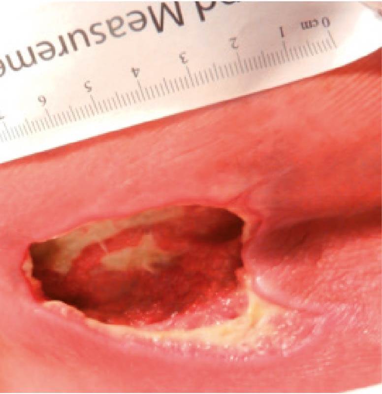
Stage 3 bedsores have the following characteristics:
- Black or rotten outer edges
- Crater-like indentation
- Dead, yellowish tissue
- Visible fat tissues
Stage 3 bedsores are quite deep, but tendons, ligaments, muscles, and/or bones will not be visible. If they are, the patient likely has a stage 4 bedsore. That said, health care providers may not be able to properly stage every severe bedsore.
Two complications may delay a stage 3 bedsore diagnosis:
- Deep tissue injuries: A deep tissue injury occurs when there is no open wound but the tissues beneath a patients skin are damaged.
- Unstageable injuries: If a doctor cannot see the base of the sore due to slough or eschar in the wound bed, they cannot make a diagnosis.
Even if a bedsore cannot be staged, doctors can still recommend treatments to start the healing process.
Also Check: What To Do For A Peptic Ulcer
How Can I Tell If I Have A Pressure Sore
- First signs. One of the first signs of a possible skin sore is a reddened, discolored or darkened area . It may feel hard and warm to the touch.
- A pressure sore has begun if you remove pressure from the reddened area for 10 to 30 minutes and the skin color does not return to normal after that time. Stay off the area and follow instructions under Stage 1, below. Find and correct the cause immediately.
- Test your skin with the blanching test: Press on the red, pink or darkened area with your finger. The area should go white remove the pressure and the area should return to red, pink or darkened color within a few seconds, indicating good blood flow. If the area stays white, then blood flow has been impaired and damage has begun.
- Dark skin may not have visible blanching even when healthy, so it is important to look for other signs of damage like color changes or hardness compared to surrounding areas.
- Warning: What you see at the skins surface is often the smallest part of the sore, and this can fool you into thinking you only have a little problem. But skin damage from pressure doesnât start at the skin surface. Pressure usually results from the blood vessels being squeezed between the skin surface and bone, so the muscles and the tissues under the skin near the bone suffer the greatest damage. Every pressure sore seen on the skin, no matter how small, should be regarded as serious because of the probable damage below the skin surface.
Recommended Reading: Healing Ulcerative Colitis With Plant Based Diet
Can Bedsores Be Prevented
Bedsores can be prevented by inspecting the skin for areas of redness every day with particular attention to bony areas. Other methods of preventing bedsores and preventing existing sores from getting worse include:
- Turning and repositioning every 2 hours
- Sitting upright and straight in a wheelchair, changing position every 15 minutes
- Providing soft padding in wheelchairs and beds to reduce pressure
- Providing good skin care by keeping the skin clean and dry
- Providing good nutrition because without enough calories, vitamins, minerals, fluids, and protein, bed sores cant heal, no matter how well you care for the sore
How The Intervention Might Work
Animal experiments conducted over 40 years ago suggested that acute wounds heal more quickly when their surfaces are kept moist rather than left to dry and scab . A moist environment is thought to provide optimal conditions for the cells involved in the healing process, as well as allowing autolytic debridement , which is thought to be an important part of the healing pathway .
The desire to maintain a moist wound environment is a key driver for the use of wound dressings and related topical agents. Whilst a moist environment at the wound site has been shown to aid the rate of epithelialisation in superficial wounds, excess moisture at the wound site can cause maceration of the surrounding skin , and it has also been suggested that dressings that permit fluid to accumulate might predispose wounds to infection . Wound treatments vary in their level of absorbency, so that a very wet wound can be treated with an absorbent dressing to draw excess moisture away and avoid skin damage, whilst a drier wound can be treated with a more occlusive dressing or a hydrogel to maintain a moist environment.
Some dressings are now also formulated with an ‘active’ ingredient .
You May Like: How To Cure Duodenal Ulcer
Nursing Home Neglect And Stage 3 Bedsores
When a lack of care causes a resident to develop a stage 3 bedsore, nursing home neglect may have occurred.
Neglect is not the same thing as making a simple, harmless mistake its a life-threatening error or series of errors. Sadly, poorly trained or inattentive staff can provide consistently poor care to residents, which can lead to bedsores.
Nursing home neglect often goes hand-in-hand with another issue: understaffing in long-term care facilities. When there are less staff members available, residents may have to wait long hours before their health care needs are addressed.
In chronically understaffed nursing homes, care problems can go unresolved for months making it more likely for bedsores to develop.
Understaffing may lead to stage 3 bedsores, as caretakers are:
- Less likely to notice or treat bedsores in their early stages
- At a greater risk of forgetting to care for every resident
- More likely to leave a resident in bed or a wheelchair for too long
Has your loved one suffered from a stage 3 bedsore?Get a free case review to get justice.
Foam Dressing For Pressure Ulcer
Foam dressing is a kind of new dressing made of foaming medical polyurethane which contains CMC.
Foam dressing:
foam dressing for pressure ulcer
Designed to promote a moist environment, thereby accelerating the healing of many difficult-to-treat wounds or chronic wounds. Substances that help to provide ideal moisture conditions include alginate fiber, foam, and hydrocolloid dressings
Hydrophilic Dressings draw exudate from the wound and protect the wound from bacterial contamination.
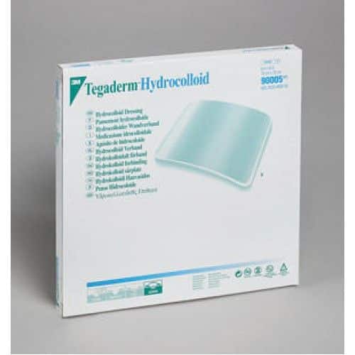
The Latex-Free Wound Dressing is ideal for Stage II, III, and IV pressure ulcers. A handy ruler is printed on each package for quick measurements.
Mechanisms:
Don’t Miss: Best Treatment For Ulcerative Colitis Flare Up
Appendix 11 Time To Event Data: Direct Evidence
The duration of followup ranged from 3 to 26 weeks, but the distribution was insufficient to allow modelling of time dependence in the network.
Seven studies reported timetoevent data. We calculated the hazard ratio using the method and spreadsheet from Tierney 2007 one study reported the hazard ratio directly, adjusted for exudate level. The timetohealing data are shown in Analysis 3.1 and summary statistics for the timetohealing and the proportion healed are compared in Table 22 for the studies that report both healing outcomes.
In the individual network, two studies in 95 participants suggested that the time to healing may have been quicker for hydrocolloid versus saline gauze there was no heterogeneity . One study in 24 participants suggested healing may have been quicker for collagenase ointment compared with hydrocolloid . In the other studies, the CI showed much uncertainty.
There was some suggestion of a time dependent effect because there were qualitative and quantitative differences between the HR and the RR: for shorter studies , the HR gave a smaller effect than the RR, but for the medium and longer term studies the HR gave a larger effect than the RR, suggesting that wounds that heal do so relatively quickly.
Analysis
Comparison 4 Direct evidence: group interventions, timetohealing data, Outcome 1 Timetohealing .