What Role Does Diet And Nutrition Play In Ulcerative Colitis
Diet does not cause the development of ulcerative colitis nor can any special diet cure the disease. However, the foods you or your child eat may play a role in managing symptoms and lengthening the time between flareups.
Some foods may make symptoms worse and should be avoided, especially during flareups. Foods that trigger symptoms are different from person to person. To narrow down what foods affect you, keep track of what you eat each day and how you feel afterward .
Problem foods often include:
- High sugar foods and drinks.
- Carbonated beverages.
- High-fiber foods.
In addition to the problem foods listed above, infants, children and teenagers can also experience issues with:
- Dairy products.
Keep a careful eye on your childs diet and nutrition. Their appetite may decrease during a flareup and they might not eat enough to stay healthy, and grow. Also, the inflammation caused by ulcerative colitis may keep their digestive tract from absorbing enough nutrients. This can also affect your childs health. For these reasons, you may have to increase the amount of calories your child consumes.
Its best to work with your provider and nutritionist to come up with a personalized diet plan if you or your child has ulcerative colitis.
Why Ct Scan Is Not The Gold Standard For Diagnosis
Dr. Gingold explains, Other things that can look similar to Crohns disease on CT scan would include cancer, appendicitis and certain infections such as Yersinia, campylobacter, salmonella Entamoeba and rarely tuberculosis.
Dr. Gingold attributes his success to the extra time he spends with his patients. His areas of expertise include reflux disease, Barretts esophagus, capsule endoscopy, chronic liver disease and inflammatory bowel disease.
Lorra Garrick has been covering medical, fitness and cybersecurity topics for many years, having written thousands of articles for print magazines and websites, including as a ghostwriter. Shes also a former ACE-certified personal trainer.
Utility Of Emergency Department Ct Scans In Patients With Ulcerative Colitis
- Dion Booras1, Danielle La Selva2 and Michael V Chiorean2*
- 1 Department Of Internal Medicine, Virginia Mason Medical Center, Seattle, Washington, United States
- 2 Division Of Gastroenterology And Hepatology, Digestive Disease Institute, Virginia Mason Medical Center, Seattle, Washington, United States
*Corresponding Author:
Tel:Received DateAccepted DateDOI:
Don’t Miss: What Dies A Stomach Ulcer Feel Like
How Do You Calm A Colitis Flare Up
Thickening Of The Bowel Wall

Thickening of the bowel wall may be caused by several pathologic conditions or be a normal variant . When thickening of the bowel wall is identified on CT, several imaging features must be assessed in order to narrow the differential diagnosis: length of involvement, degree of thickening, symmetric versus asymmetric involvement, pattern of attenuation and perienteric abnormalities . Each of these features may have a different significance according to the acute or chronic onset of clinical symptoms and will be further discussed in an algorithm approach .
Also Check: Preventing Pressure Ulcers In Nursing Homes
Who Diagnoses Ulcerative Colitis
If you have symptoms of ulcerative colitis, your regular healthcare provider will probably refer you to a specialist. A gastroenterologist a doctor who specializes in the digestive system should oversee the care for adults. For young patients, a pediatric gastroenterologist who specializes in children should manage the care.
How Often Do I Need A Colonoscopy
Especially when you have symptoms or are just starting or changing medications, your doctor may want to periodically look at the inside of the rectum and colon to make sure the treatments are working and the lining is healing. How often this is needed is different for each person.
Ulcerative colitis also increases your chance of developing colon cancer. To look for early cancer signs, your healthcare provider may have you come in for a colonoscopy every one to three years.
Read Also: Colon Cleanse For Ulcerative Colitis
Definition Of The Etiologies Of Acute Colitis
The episode of acute colitis was considered as infectious if either the routine BD-Max or the multiarray PCR assay FilmArray GI panel was positive. Escherichia coli spp. EAEC, EPEC and ETEC identified only by FilmArray GI panel were considered as carriage due to ongoing doubts regarding their pathogenicity. Pathogens were further identified by fecal culture. If BD-Max was negative, diagnosis was obtained using colonoscopy with or without biopsies. In non-infected cases, we defined the etiology of colitis as ischemic, or secondary to colorectal cancer or IBD in the presence of suggestive macroscopic aspect and/or histopathology. Patients later registered in the Swiss IBD cohort were considered to have IBD. To avoid missing a secondary diagnosis in patients with infectious etiology, medical files were reviewed at the date of the inclusion of the last patient to rule out later diagnosis of IBD and/or colorectal cancer. Undetermined colitis, in which the etiology of acute colitis was unknown, was defined as normal colonoscopy and, if available, normal histology.
B Patientlevel Colitis Diagnosis Performance
An SVM classifier was trained for patientlevel diagnosis. The training set and testing set are consistent with the data used for colitis detection. Figure 11 shows the patientlevel diagnosis test ROC curves for the RCNN and Faster RCNN methods, respectively. The area under the ROC curve was 0.978 ± 0.009 with the RCNN method, and it improved to 0.984 ± 0.008 with the Faster RCNN method . At the optimal operating point , the RCNN method correctly identified 90.4% of the colitis patients and 94.0% of normal cases. The sensitivity improved to 91.6% and the specificity was improved to 95.0% at the optimal operating point for the Faster RCNN method. ZF net was used in both methods presented in the figures.
Patientlevel diagnosis ROC curves of RCNN and Faster RCNN.
Figure 12 shows two examples of normal cases that were misdiagnosed as patients with colitis. The stomach and normal colon were identified as colitis in these two normal cases. These regions are misdetected as colitis because the gastric wall thickness can be greater than 3 mm if the stomach is not distended and because residual fecal material in normal colon was misinterpreted as abnormal wall thickening.
Examples of false colitis diagnoses in two normal patients. The appearances of stomach and normal undistended colon were similar to colitis.
Also Check: Managing Ulcerative Colitis Flare Ups
Can You See Bowel Inflammation On A Ct
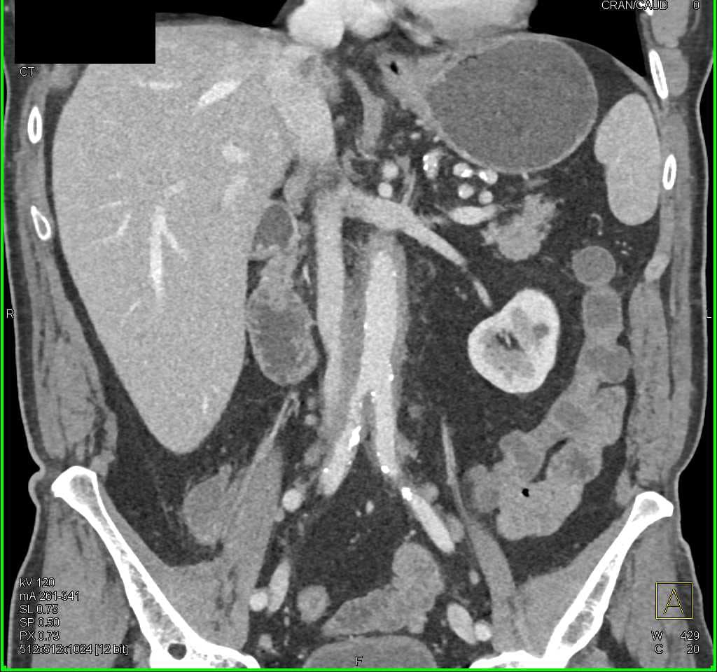
A stricture, also known as an obstruction, is a narrowing of the small or large intestine that can be discovered by CT scans of the gastrointestinal tract. Furthermore, the test may reveal inflammation in the small intestine, which could indicate that you are suffering from Crohns disease.
A CT scan can determine whether a diverticula is inflamed, the bowel wall is inflamed, or a fatty strand is present in the colon. CT provides better quality of care and a more accurate diagnosis by visualizing peritoneum and colonic complications, both of which are difficult to detect with conventional methods. CT scans can be used to diagnose Crohns disease and its complications. The sooner complications are identified, the better the patient will receive care.
How Upper Gi X
X-rays can also give your doctor clues to diagnose IBDs. Upper gastrointestinal tract radiography, also known as an upper GI series, uses a form of X-ray called fluoroscopy and a contrast material, such as barium. Fluoroscopy allows your doctor to see how organs function while in motion. An upper GI series shows the esophagus, stomach, and small intestine, with the barium coating giving your doctor a clearer picture of the function and anatomy of these organs.
If you are having symptoms of Crohns disease, ulcerative colitis, or another IBD and your doctor has ordered imaging, the experts at American Health Imaging can help. We offer reliable, cost-effective imaging performed by expert radiologists. Request an appointment at the location closest to you.
Read Also: How To Treat Skin Ulcer On Leg
Management Of Patients With Computed Tomography
Patients suffering from CT-proven acute colitis and compatible symptoms were routinely hospitalized. Blood tests were collected at admission and then according to clinical judgement. First feces were collected in Cary-Blair medium and sent for microbiological analyses using BD-Max. Additional fecal samples were collected and stored in a 4 °C fridge for a maximum of 24 h, before being transferred to a 80 °C freezer. These samples were sent to an external laboratory for calprotectin determination. Additional microbiological analyses were performed in parallel using FilmArray GI panel in all patients. Antibiotic treatment with intravenous 2g ceftriaxone 1×/day and oral 500mg metronidazole 3×/day was initiated from admission and continued for at least 5 days, according to institutional guidelines. Then, antibiotics treatment was continued per os using 500mg ciprofloxacine 2×/day and 500mg metronidazole 3×/day for a total of 10 days in case of episode of undetermined etiology, or adapted to microbiological results from the fecal samples.
Idiopathic Inflammatory Bowel Disease

Bowel wall thickening with a stratified pattern may be also seen in both ulcerative colitis and Crohns disease, indicating acute, active disease .
Crohns disease may occur in any part of the gastrointestinal tract but predominantly affects the small bowel, particularly the ileum and right colon . CT signs favouring Crohns disease include discontinuous involvement of the bowel wall , prominent vasa recta and signs of transmural inflammation such as fistulas and abscesses, and proliferation of the fat along the mesenteric border of the bowel .
Fig. 13
Stratified appearance in Crohns disease. Axial contrast-enhanced CT scan of the abdomen shows concentric wall thickening of small bowel loops with a stratified appearance indicating active disease . Also note a fistula connecting the bowel loops, a common finding in Crohns disease
Don’t Miss: What Do Venous Leg Ulcers Look Like
V Describe The Advantages And Disadvantages Of The Alternative Techniques For Diagnosis Of Ulcerative Colitis
Acute abdominal series x-rays
-
Can provide quick information regarding bowel dilation, obstruction and possible free air.
Disadvantages
-
Limited diagnostic utility for ulcerative colitis
Barium enemas
-
Can show mucosal irregularity, benign stricturing, and haustral changes.
Disadvantages
-
Limited utility in surveillance of ulcerative colitis.
Can A Ct Scan Detect Problems In The Colon
A computed tomography scan can be used to detect colorectal cancer, identify cancer in other areas of the body, and evaluate the effectiveness of cancer treatment.
An x-ray of the large intestine is performed using computed tomography colonography or virtual colonoscopy, and cancers and growths known as polyps are evaluated. The goal of CT colonography is to obtain a detailed image of the internal organs using low dose radiation CT scanning. The only previous treatment was less invasive and involved the doctor inserting an endoscope into the rectum and passing it through the colon. Women and men are encouraged to begin colon cancer screenings as soon as they reach the age of 45, according to the American Cancer Society. CT colonography can be performed once every five years, according to the American Cancer Society. Colorectal cancer is distinguished by persistent changes in bowel habits, the presence of blood in the stool, abdominal discomfort, and pain. Radiation from the body is carried by X-rays, which are radiations that come in forms such as light or radio waves.
Don’t Miss: Can Ulcers Give You Headaches
Ct Scans For Ibd Diagnosis
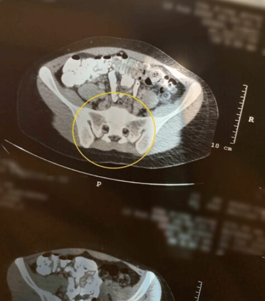
Computed tomography, or CT scans, use special X-ray equipment to take cross-sectional pictures of structures inside the body. Your doctor can use a pelvic CT scan to view images of your gastrointestinal tract and abdominal organs to look for signs of Crohns disease or ulcerative colitis, such as inflammation, and determine where the inflammation occurs.
CT enterography is a special type of CT scan that uses contrast material to better show the small intestine and additional structures in the abdomen and pelvis. This scan can show whether you have ulcerative colitis or Crohns disease and the location and severity of the disease.
Can You See Crohn Disease On A Ct Scan
CT and MR imaging are commonly used to detect Crohns disease, but their sensitivity can be limited. For patients who are at an early stage of disease, colonoscopy and enteroclysis studies are recommended.
Using a risk stratification model, it was determined whether or not to have CT scans performed in Crohns disease patients. A model cut emergency department scans of these patients by 43%, and the missed rate was only 1.8%. The United States could prevent over 250 cancer cases by treating them at emergency departments and saving more than $80 million per decade. Complications were caused by only C-reactive protein and erythrocyte sedimentation rate levels. In 18.5% of cases, a CT scan would be avoided if the ESR was 5 x CR and the value of the CT scan was 10 or less. Scans could, however, be avoided by using a more complex logistic regression model instead of the simple equation.
Read Also: Natural Ways To Cure Stomach Ulcers
What Causes Ulcerative Colitis Flareups
When youre in remission from ulcerative colitis, youll want to do everything you can to prevent a flareup. Things that may cause a flareup include:
- Emotional stress: Get at least seven hours of sleep a night, exercise regularly and find healthy ways to relieve stress, such as meditation.
- NSAID use: For pain relief or a fever, use acetaminophen instead of NSAIDs like Motrin® and Advil®.
- Antibiotics: Let your healthcare provider know if antibiotics trigger your symptoms.
Why Would A Doctor Order A Ct Scan Of The Abdomen
Doctors may use an abdominal CT scan to look for signs of injury, infection, or disease in organs such as the colon, spleen, liver, or kidneys. A CT scan usually takes only a few minutes. The procedure does not hurt, but some people may find it uncomfortable to lie still for the duration of the scan.
Read Also: Best Medication For Stomach Ulcers
What Can I Expect If I Have A Diagnosis Of Ulcerative Colitis
Ulcerative colitis is a lifelong condition that can have mild to severe symptoms. For most people, the symptoms come and go. Some people have just one episode and recover. A few others develop a nonstop form that rapidly advances. In up to 30% of people, the disease spreads from the rectum to the colon. When both the rectum and colon are affected, ulcerative symptoms can be worse and happen more often.
You may be able to manage the disease with medications. But surgery to remove your colon and rectum is the only cure. About 30% of people with ulcerative colitis need surgery.
What Gets Stored In A Cookie

This site stores nothing other than an automatically generated session ID in the cookie no other information is captured.
In general, only the information that you provide, or the choices you make while visiting a web site, can be stored in a cookie. For example, the site cannot determine your email name unless you choose to type it. Allowing a website to create a cookie does not give that or any other site access to the rest of your computer, and only the site that created the cookie can read it.
You May Like: Difference Between Ulcerative Colitis And Diverticulitis
How Is Ulcerative Colitis Treated
Theres no cure for ulcerative colitis, but treatments can calm the inflammation, help you feel better and get you back to your daily activities. Treatment also depends on the severity and the individual, so treatment depends on each persons needs. Usually, healthcare providers manage the disease with medications. If your tests reveal infections that are causing problems, your healthcare provider will treat those underlying conditions and see if that helps.
The goal of medication is to induce and maintain remission, and to improve the quality of life for people with ulcerative colitis. Healthcare providers use several types of medications to calm inflammation in your large intestine. Reducing the swelling and irritation lets the tissue heal. It can also relieve your symptoms so you have less pain and less diarrhea. For children, teenagers and adults, your provider may recommend:
Children and young teenagers are prescribed the same medications. In addition to medications, some doctors also recommend that children take vitamins to get the nutrients they need for health and growth that they may not have gotten through food due to the effects of the disease on the bowel. Ask your healthcare provider for specific advice about the need for vitamin supplementation for your child.
You might need surgery that removes your colon and rectum to:
- Avoid medication side effects.
- Prevent or treat colon cancer .
- Eliminate life-threatening complications such as bleeding.
What Causes Ulcerative Colitis
Researchers think the cause of ulcerative colitis is complex and involves many factors. They think its probably the result of an overactive immune response. The immune systems job is to protect the body from germs and other dangerous substances. But, sometimes your immune system mistakenly attacks your body, which causes inflammation and tissue damage.
Don’t Miss: To Prevent Pressure Ulcers You Must
Similar Articles Being Viewed By Others
Carousel with three slides shown at a time. Use the Previous and Next buttons to navigate three slides at a time, or the slide dot buttons at the end to jump three slides at a time.
10 August 2021
Yuko Akazawa, Tomohito Morisaki, Fuminao Takeshima
19 January 2021
Sarah E. R. Bailey, Gary A. Abel, Willie Hamilton
18 June 2019
Eamonn M. M. Quigley
27 May 2021
Eriko Yasutomi, Toshihiro Inokuchi, Hiroyuki Okada
volume 12, Article number: 9730
Approach To The Thickened Bowel Wall
When thickening of the small or large bowel wall is identified on CT, the first step to take is to access the extent of the involved bowel. Distinction should be made between focal and segmental or diffuse involvement . This is an important step in differentiating between benign and malignant causes of bowel wall thickening: while most bowel tumours present as a focal involvement, segmental and diffuse thickening of the bowel wall are usually caused by benign conditions . The exception is a small bowel lymphoma, which typically shows as a segmental distribution .
Fig. 1
Algorithm approach to the bowel wall thickening. CD Crohns disease, TB tuberculosis, IBD inflammatory bowel disease, RE radiation enteritis. Adapted from the electronic poster Bowel wall thickeninga complex subject made simple DOI:10.5444/esgar2011/EE-063
You May Like: Can You Have An Ulcer In Your Colon