Why Is Duoderm Used To Treat Bed Sores
DuoDERM dressing bandages successfully block oxygen, bacteria, and water vapor from permeating the bandage. DuoDERM has become incredibly popular due to it being a wound dressing that doesn’t stick to the wound. DuoDERM is also a wound care treatment that can be easily removed without injuring the skin underneath.
DuoDERM wound dressing bandages are ideal for those wounds that are experiencing mild to moderate seepage. A DuoDERM dressing can be used on all types of bedsores, including stage 1, 2, 3, and 4.
A DuoDERM dressing can be used on a wound for several days. However, the exact time period during which a DuoDERM dressing will be effective will depend largely on the moisture that is present within the wound. In most cases, a DuoDERM dressing can remain on a wound for between three and seven days. One of the benefits of choosing DuoDERM is that it is ideal for wounds that are wet and will remain in place even in a moist environment.
Since a DuoDERM patch is able to create a moisture barrier, it’s important to always make sure that the bedsores are not infected, as a DuoDERM dressing would only make the problem worse. As long as infection is not present, a DuoDERM patch will normally cause a wound to begin smelling strongly after a couple of days without any cause for concern. Again, due to the containment of healthy moisture within the DuoDERM patch, the development of a smell can be expected.
Management Of Sacral Ulcers Varies By Ulcer Stage
It is important to properly stage pressure ulcers for several reasons, but two of the most important are for prognosis and management planning. Stage 1 and stage 2 pressure ulcers heal by regenerating tissue in the wound. Stage 3 and stage 4 pressure ulcers, on the other hand, heal through scar formation, which means the borders of the wound contract as it heals.
For Stage 1 sacral ulcers, the primary goal of therapy is to ensure adequate tissue perfusion and to protect the wound from further damage.2 This means preventing the sacrum from chronically squeezing the skin and preventing blood flow to the area. The goal of therapy for Stage 2 ulcers is to encourage tissue regeneration and protect the wound surface. For stage 3 or 4 ulcers, management efforts are focused on promoting tissue granulation and epithelialization.
Who Is Most At Risk For Bedsores
Since the occurrence of bedsores requires prolonged pressure to a particular area of the skin, they are most common for those who remain in bed or on bedrest for an extended period of time. Typically, those with certain medical conditions that limit their ability to move or change positions are the most likely to be affected. However, bedsores can also occur when individuals spend the majority of their time in bed or even in a chair.
Unfortunately, bedsores can occur quite quickly. While there are several treatments and wound care treatment options available to address bedsores once they occur, it’s always best to try and prevent them from occurring in the first place. If bedrest is required or if motion is limited, try to change positions as much as possible and pay close attention to the skin and take note of any changes to its coloring or texture.
Don’t Miss: Is Okra Good For Ulcerative Colitis
Cost And Quality Of Life Data
Cost and quality of life data used in the studies were also generally poor quality or lacking. When quality of life measures were reported they tended to be linear analogue scales or simple Likert-type scales. The inclusion of more sophisticated measures of quality of life when evaluating dressings is an area that needs to be tackled. This is particularly important as it may be one of the few ways to distinguish between dressings. The impact of venous ulcers on quality of life has been studied, but within randomised controlled trials quality of life data were very poor or omitted altogether.
The poor reporting of cost data was a particular concern. Where such data were collected,w7w14w29 the reporting did not conform to rigorous guidelines for economic evaluations. The trials simply totalled the monetary cost of the dressings and did not examine their cost effectiveness. This was illustrated in the hydrocolloid versus alginate comparison, where costs were reported for the interventions but insufficient detail was provided on their derivation.
Collagen Vs Hydrocolloid In Treatment Of Pressure Ulcers

Am Fam Physician. 2003 Aug 15 68:742-743.
Effective wound healing and cost considerations dictate the dressing choice in the treatment of pressure ulcers. Graumlich and colleagues performed a study to compare hydrocolloid dressings with collagen in the healing of stage II and III pressure ulcers. Hydrocolloid is a moist, vapor-permeable, occlusive dressing used in wound healing. Collagen, extracted from bovine skin, enhances wound healing through a variety of probable mechanisms, such as angiogenesis, epithelialization, and granulation.
The investigators enrolled nursing home patients with stage II to III pressure ulcers in a single-blind, randomized trial, in which participants received collagen or hydrocolloid dressings, continuing treatment for eight weeks. The definition of complete healing was 100 percent epithelial coverage of the study ulcer, with the proportion of completely healed ulcers serving as the primary end point. Blinded observers used validated, standardized techniques to record ulcer length, width, and appearance.
There was a nonsignificant trend favoring collagen for healing ulcers deeper than 2 mm at baseline. Collagen treatment was more expensive and required more nursing interventions per week. The number needed to treat with collagen is 70 patients for up to eight weeks before one healing event will occur in patients who would otherwise receive hydrocolloid treatment.
Read the full article.
You May Like: Remicade Vs Entyvio For Ulcerative Colitis
What Kind Of Dressings Are Used For Pressure Ulcers
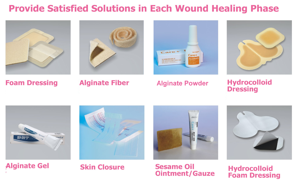
Foam Dressings . Stage III Pressure Ulcer. Open wound that breaks through all skin levels Visible fat layer, but not bone, tendon or muscle Wound may begin expanding under adjacent intact skin Foam Dressings . Hydrogels . Hydrocolloids. Alginate Dressings. Stage IV Pressure Ulcer. Open wound that exposes bone, tendon or muscle
What Are Some Of The Signs And Symptoms Of Bed Sores
As mentioned, bedsores can arise quite quickly. However, in most cases, there are some warning signs that you can keep an eye out for. Doing so may be able to help you reduce the severity of the bedsores or prevent them from occurring completely. Some of the most common symptoms of pressure ulcers include:
- Changes to the color or texture of the skin
- Swelling of the skin
- Pus-like draining of the skin
- Parts of the skin that feel either colder or warmer than the rest
- Skin that is tender to the touch
There are several stages of bedsores, each of which is defined based upon the severity, depth, and additional characteristics of the wound. The extent of damage and injury can vary greatly, ranging from slightly red skin to deep injury that affects the skin as well as the muscle and even bone underneath.
Also Check: What Foods To Eat With Ulcerative Colitis
What Type Of Dressing Is Used On A Stage 1 Pressure Ulcer
Wound dressings for a grade 1 pressure ulcer should be simple and offer protection without risking any further skin damage, especially if the patient is sliding down the bed or chair causing the dressing to ruck. A film dressing or a thin hydrocolloid would be appropriate to protect the wound area.
Pressure Ulcer Prevention And Treatment: Use Of Prophylactic Dressings
Accepted for publication 5 March 2015
11 October 2016Volume 2016:3 Pages 117121
Kathleen Reid,1 Elizabeth A Ayello,2 Afsaneh Alavi,3
Introduction
Pressure ulcers are a major cause of mortality, morbidity, patient suffering, and cost on the health care system worldwide. The management of pressure ulcers is a compounding challenge to health care professionals across disciplines. Individuals who acquire pressure ulcers often require long-term interventions, representing a large economic burden to the health care system. It has been estimated that in Australia, these injuries increase the length of hospital stay and subsequently incur $285 million in cost annually.1 Since 2008, the Centers for Medicare and Medicaid Services no longer reimburses American hospitals at a higher rate for any pressure ulcer that occurs during a patients hospitalization, which provides a strong financial stimulus for pressure ulcer prevention protocols to be implemented.2 Indeed, the profound impact of pressure ulcers on the emotional, physical, mental, and social domains of life has been shown in different studies.3 Current management strategies target pressure-relieving surfaces: patient repositioning, nutritional support, and application of protective dressings to prevent pressure injuries. Dressings are accessible and easily implemented devices however, they can also contribute to high health care costs. Therefore, it is important to evaluate their efficacy.
Prophylactic role of dressings
Read Also: Is Ulcerative Colitis Considered An Autoimmune Disease
Thin Flexible And Versatile
Designed to reduce the risk of further skin breakdown due to friction.
DuoDERM® Extra Thin Dressing can be used as a primary hydrocolloid dressing for dry to lightly exuding wounds.
It can be used as a secondary dressing to secure an AQUACEL® Dressing or an AQUACEL® Ag Dressing.
The European Pressure Ulcer Advisory Panel and The National Pressure Ulcer Advisory Panel guidelines recommend the usage of hydrocolloids for the management of pressure ulcers.1
DuoDERM® Extra Thin Dressing can be used to manage stage I and stage II pressure ulcers.
Box : Comparisons Of Dressing Types
Hydrocolloids
-
Versus simple/non-adherent dressings
-
Versus other hydrogel dressings
The primary outcome measure was time to complete ulcer healing or proportion of ulcers completely healed. We excluded composite outcome measures such as ânumber of ulcers healed or improved.â
We identified randomised controlled trials by searching Medline, Embase, and CINAHL, as well as the Cochrane Wounds Group specialised trials register up to April 2006. Box 2 shows details of all the databases searched and search terms used. We also sought grey literature by examining conference proceedings. We placed no restrictions in terms of language or year of publication. We also hand searched key journals, checked citations, and contacted experts in the field of wound care to enquire about ongoing and recently published trials.
Don’t Miss: Does Ulcerative Colitis Cause Fever
Hydrogel Dressings For Treating Pressure Ulcers
Background
Pressure ulcers, also known as bedsores, decubitus ulcers and pressure injuries, are areas of injury to the skin or the underlying tissue, or both. Pressure ulcers can be painful, may become infected, and affect quality of life. Those at risk of pressure ulcers include those with spinal cord injuries and people who are immobile or who have limited mobility such as some elderly people and people with acute or chronic conditions. In 2004 the total annual cost of treating pressure ulcers in the UK was estimated as being GBP 1.4 to 2.1 billion, which was equivalent to 4% of the total NHS expenditure. Pressure ulcers have been shown to increase length of hospital stay and the associated hospital costs. Figures from the USA suggest that ‘pressure ulcer’ was noted as a diagnosis for half a million hospital stays in 2006 for adults, the total hospital costs of these stays was USD 11 billion.
Dressings are one treatment option for pressure ulcers. There are many types of dressings that can be used these can vary considerably in cost. Hydrogel dressings are one type of available dressing. Hydrogel dressings contain a large amount of water that keeps ulcers moist rather than letting them become dry. Moist wounds are thought to heal more quickly than dry wounds. In this study we investigated whether there is any evidence that pressure ulcers treated with hydrogel dressings heal more quickly than those treated with other types of dressings or skin surface treatments.
How Do You Apply A Duoderm Dressing

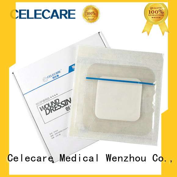
One of the big advantages of the DuoDERM patch is that it is easy to apply and easy to remove. The clear backing of the DuoDERM dressing allows for ease of placement as well as monitoring of the wound during the healing process. Not only this but the tapered edge of the DuoDERM patch is able to contour to just about any difficult area of the body.
When applying the wound dressing, you’ll want to:
- Make sure your hands have been thoroughly cleaned.
- Clean the wound with a saline solution.
- Remove the dressing from the sleeve.
- Peel back the release paper as well as the margin tabs.
- Apply the wound care treatment.
- Ensure good contact with the skin on all sides. You can make sure this is done properly by taking care to smooth any folds.
When in doubt, don’t hesitate to look at the packaging for your wound care treatment to ensure that you are using the optimal method of applying and securing your hydrocolloid bandages.
Also Check: Diet Plan For Ulcerative Colitis Flare Up
Which Wound Dressing Is Best For Your Pressure Ulcer
Now that weve touched on some of the more common types of dressings used for pressure ulcers, you may be wondering which is the best for your particular situation. The answer will depend on multiple factors including where the pressure ulcer is located, how severe the bedsore is, and the degree of skin and tissue damage. Talk to your health care professional about any pressure wounds you notice on your body as soon as possible.
How The Intervention Might Work
The principle of moist wound healing governs wound care practice today. This is optimised through the application of occlusive or semiocclusive dressings and preparation of the wound bed . Animal experiments performed 50 years ago suggested that acute wounds healed more quickly when their surface was kept moist, rather than being left to dry and to scab . examined the rate of epithelialisation in experimental wounds cut into the skin of healthy pigs, comparing wounds with a natural scab exposed to the air against wounds that were covered with polythene film. He found that epithelialisation occurred more quickly in the latter.
The principle of moist wound healing has led to the development of several commercially available wound dressings to support optimal healing processes. These have revolutionised wound management products include hydrogels that retain moisture in contact with the wound, hydrocolloids that absorb small amounts of excess moisture without drying the wound bed, absorbent foams, alginates, adhesive dressings, nonadhesive dressings and siliconebased lowadherent dressings. Hydrocolloid dressings are composed of a layer of sodium carboxymethylcellulose bonded onto a vapourpermeable film or foam pad. These occlusive dressings absorb exudate whilst maintaining a moist wound environment. Fibrous hydrocolloids are a subset of dressings that are designed for use in wounds with heavy exudate in lieu of alternate dressing types such as alginates .
You May Like: Do Ulcers Cause Weight Loss
The Efficacy Of Duoderm
A study by Michel Hermans treating small partial-thickness burns found that “HydroColloid Dressings provide an optimum wound environment for more rapid re-epithelialization than either allografts or SSD. The cosmetic and functional results were also excellent.”1 A meta-analysis of pressure ulcers performed by Matthew Bradley comparing a hydrocolloid dressing with a traditional treatment suggested that treatment with the hydrocolloid resulted in a statistically significant improvement in the rate of healing compared to wet-to-dry dressings.2
Disadvantages Of Hydrocolloid Dressings
These types of dressings are not appropriate for all wounds and should not be used if there is heavy exudate or infection. Other disadvantages include:
- It can be difficult to assess the wound through the bandage
- Bandages might curl or roll on edges
- Sometimes dressing adheres to the wound and causes trauma to the fragile skin when removed
- Dressings can cause periwound maceration or hypergranulation of wound
Read Also: Does Stelara Work For Ulcerative Colitis
Removing A Hydrocolloid Dressing
Follow these procedures to remove a hydrocolloid dressing from a wound:
How To Apply Hydrocolloid Dressings
Applying a hydrocolloid dressing is similar to the best practices for most wound care. Follow these steps:
Also Check: Medical Management Of Ulcerative Colitis
External Validity Of Included Trials
The external validity of many of these trials is threatened by the fact that they limited inclusion by ulcer size. Of the 23 studies that reported details of baseline ulcer area, 15 included only ulcers of less than 10 cm2 and eight included only ulcers of greater than 10 cm2. Only one trial used life table analysis, summarising both how many people’s ulcers healed and how quickly they healed.w9 Many studies used rate of reduction in ulcer area as an outcome measure however, this is not necessarily a predictor of healing, particularly when used over a short period. In addition, the use of change in wound area raises questions of validity, especially when initial ulcer size varies, as the percentage change will be greater for smaller wounds. The use of rates of reduction in area can therefore be misleading.
Other outcome measures used included patient derived and nurse derived subjective measures such as âsatisfactionâ and pain. The use of subjective outcomes in trials can lead to bias, especially if the tools used are not tested for reliability and validity and if blinding to treatment allocation is not used. Bias can result from subconscious preferences of treatments by the assessors, patients, or both, or selective reporting of positive outcomes.