Prevention Of Ulcer Recurrence
Factors that are associated with ulcer nonhealing and recurrence: Overweight body mass index, history of deep venous thrombosis, large ulcer area, noncompliance with compression therapy, and triple-system venous disease involving superficial, perforating, and deep veins . The strategies to prevent the ulcer recurrence should target these factors. These could be implemented as regular clinical evaluations, patient education and life-long compression therapies. Patientâs education should be regarding skin care, elevation of the affected limb when immobile, compliance to compression therapy, encourage mobility, and exercise. To encourage, early self-referral at signs of possible skin breach.
Compression therapy
Use of compression stockings reduces ulcer recurrence and is thus highly recommended in patients of venous leg ulcers. Patients are encouraged to wear the strongest compression they can tolerate for life-long, if not contraindicated otherwise .
Infection Of Venous Leg Ulcers
The most common complication of venous leg ulcers is infection, which can arise from a variety of sources, including the following:1
- Bacterial: furuncles, ecthyma, mycobacterioses, syphilis, erysipelas, anthrax, diphtheria, chronic vegetative pyoderma, tropical ulcer
- Viral: herpes, variola virus infection, cytomegalovirus infection
- Fungal: sporotrichosis, histoplasmosis, blastomycosis, coccidioidomycosis
- Protozoal: leishmaniasis
When infection is present in the VLU, the patient may present with fever, wound fluid rich in leukocytes, increasing pain, cellulitis, necrotic tissue, and purulent exudate with or without odor. When signs of infection are present, the patient should be treated with appropriate systemic antibiotics, topical antimicrobials, or antiseptics.2 However, VLUs that are critically colonized with bacteria or bacterial biofilms without signs of systemic infection may be treated in multiple ways, including topical antibiotics. Sequential, aggressive debridement can also be used to treat infected ulcers.1
What Causes A Leg Ulcer
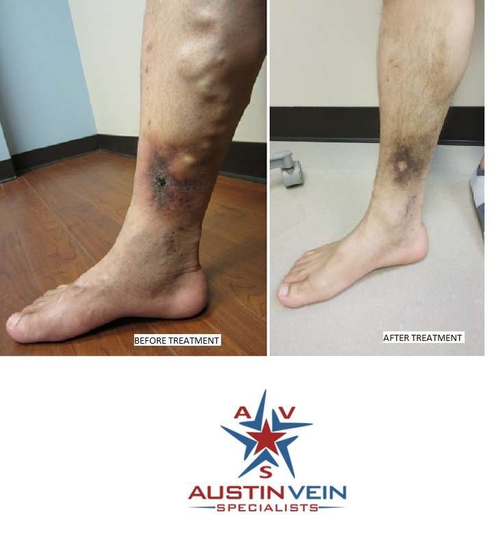
A venous leg ulcer happens when someone with poor blood circulation suffers a minor injury.
Poor blood circulation in the legs causes the pressure inside the veins to increase. Prolonged high pressure will damage the small blood vessels in the legs, making them very delicate. If the skin is subsequently broken due to even a small knock or scratch, the skin will break, creating a sore. Because of the poor circulation, the sore will take a very long time to heal. A chronic sore on the legs is called a leg ulcer.
As mentioned above, old age significantly increases the risk of a leg ulcer. Other risk factors include:
- Obesity
- History of leg injury e.g. fractured bone
- History of leg surgery e.g. knee replacement
You May Like: Nanda Nursing Diagnosis For Ulcerative Colitis
Antibiotics For Wound Infection
Jennifer Nelson
Jennifer Nelson
Jennifer is a contributing health writer who has been researching and writing health content with PlushCare for 3 years. She is passionate about bringing accessible healthcare and mental health services to people everywhere.
Dr. Katalin Karolyi
Dr. Katalin Karolyi
Katalin Karolyi, M.D. earned her medical degree at the University of Debrecen. After completing her residency program in pathology at the Kenezy Hospital, she obtained a postdoctoral position at Sanford Burnham Prebys Medical Discovery Institute, Orlando, Florida.
Read Also: Best Antibiotic For Intestinal Infection
Cleaning And Dressing The Ulcer

The first step is to remove any debris or dead tissue from the ulcer and apply an appropriate dressing. This provides the best conditions for the ulcer to heal.
A simple non-sticky dressing will be used to dress your ulcer. This usually needs to be changed once a week.
Many people find they can manage cleaning and dressing their own ulcer under the supervision of a nurse.
You May Like: What To Do For Ulcer Pain
Iodine And Silver Preparations:
Several new formulations of older topical agents should be considered to treat nonhealing wounds with or without evidence of clinical infection . Iodine, while toxic in high concentrations to tissue in vitro, can be beneficial at low doses. Cadexomer iodine releases low levels slowly into wounds and has been shown to be safe and effective at decreasing bacterial burden in the superficial compartment. Cadexomer iodine is available as an ointment and as an impregnated gauze dressing.
Silver preparations have been used on ulcers for many years. Nanocrystalline silver can deliver topical concentrations to the superficial compartment that are effective against a range of organisms, including yeast., Use of iodine and silver-containing preparations is summarized in .
Epidemiology And Risk Factors
Pressure ulcers are areas of necrosis caused by compression between bony prominences and external surfaces. The damage may be relatively minor, or it may lead to massive destruction of deeper tissues, which can cause significant morbidity and mortality. The incidence and prevalence of pressure ulcers depends on the definition of pressure ulcers used and the patient population studied. The National Pressure Ulcer Advisory Panel has classified pressure ulcers according to 4 stages :
Stage I: Nonblanchable erythema of intact skin.
Stage II: Partial-thickness skin loss involving the epidermis or dermis lesions may present as an abrasion, blister, or superficial ulcer.
Stage III: Full-thickness skin loss that may extend to, but not through, the fascia the ulcer may be undermined.
Stage IV: Full-thickness skin loss involving deeper structures, such as muscle, bone, or joint structures.
Risk factors for the development of pressure ulcers are either intrinsic or extrinsic. Limited mobility and poor nutrition are the strongest intrinsic predictors of pressure ulcer formation. Incontinence, increased age, diabetes mellitus, stroke, white race, skin abnormalities, and male sex have also been implicated by multivariate analysis in some studies . Extrinsic factors include pressure, friction, shear stress, and moisture of these, the most important is pressure.
Recommended Reading: Bacterial Vaginosis Caused By Antibiotics
Don’t Miss: Best Diet For Gerd And Ulcers
Diagnosis Of Leg Ulcers
- examine the wound
- perform some tests to measure the blood flow in your lower leg, such as the ankle-brachial index. This test compares blood pressure readings taken at the ankle and at the arm using a device called a Doppler machine
- recommend an angiogram for an arterial ulcer, to find out if the artery needs surgery to clear the blockage.
Read Also: What To Eat To Help Stomach Ulcers
F Grading The Evidence For Each Key Question
At the completion of our review, at least two reviewers will independently assign evidence grades. Conflicts will be resolved through consensus or third-party adjudication. We will grade the strength of evidence based on the quantity, quality, and consistency of the best available evidence, addressing KQs 1, 2, and 3 by adapting an evidence grading scheme recommended in the Methods Guide.13 We will apply evidence grades to the bodies of evidence about each intervention comparison for each outcome. We will assess the risk of bias of individual studies according to study design characteristics, such as confounding and selection and information biases. We will assess the strength of the best available evidence by assessing the limitations to individual study quality , consistency, directness, precision, publication bias, and the magnitude of the effect.
We will classify evidence pertaining to the KQs into four basic categories: 1) high grade 2) moderate grade 3) low grade and 4) insufficient grade .
Recommended Reading: Do Stomach Ulcers Cause Nausea
How Should I Treat A Leg Infection
The most appropriate treatment for a leg infection depends on the particular type of infection. Some affect a persons skin cells while others may involve the bone or lymph nodes. If a person suspects he has an infected leg, he should see a doctor for a diagnosis and treatment recommendation. Some seemingly minor infections can become severe if left untreated or if theyre treated with the wrong types of medication.
One type of infection that may affect a persons leg is called cellulitis. This infection involves the cells right below the skins surface, causing inflammation in the affected area. It develops when the skin is broken, such as because of a cut, broken blister or animal bite. The broken skin allows bacteria to enter the body, which leads to the infection. There are many types of bacteria that cause cellulitis, but those in the strep family are the most common.
Oral antibiotics are usually used to treat a cellulitis leg infection and usually cure it within a week or so. A patient may return to his doctor after about a week to check whether it has gone away entirely. In a severe case, a person may need to be hospitalized and treated with intravenous antibiotics.
Dont Miss: Will Doctors Prescribe Antibiotics Over Phone
Synthesis Of Ldh Doxycycline/ldh And Amoxicillin/ldh
Mg 2.6H2O and Al 3.9H2O were dissolved in 100mL distilled water. Sodium hydroxide was added dropwise until complete precipitation at pH 10.0. The precipitate suspension was stirred for 20h at 65°C, filtered, washed several times with double-distilled water and finally dried at 40°C.
Doxycycline-LDH was synthesized by repeating the same procedures and adding 0.005mol of doxycycline to the medium before precipitation. The doxycycline-LDH-precipitate suspension was stirred at room temperature for 20h, filtered, washed, and dried at 40°C. Amoxicillin-LDH was synthesized by adding a solution of amoxicillin to Mg: AL LDH for 24h at room temperature. The precipitate was filtered, washed and dried at 40°C. All steps are shown in Scheme .
Also Check: Doctors Specializing In Ulcerative Colitis
Cleveland Clinic Heart Vascular & Thoracic Institute Vascular Medicine Specialists And Surgeons
Choosing a doctor to treat your vascular disease depends on where you are in your diagnosis and treatment. The following Heart, Vascular & Thoracic Institute Sections and Departments treat patients with all types of vascular disease, including blood clotting disorders:
Section of Vascular Medicine: for evaluation, medical management or interventional procedures to treat vascular disease. In addition, the Non-Invasive Laboratory includes state-of-the art computerized imaging equipment to assist in diagnosing vascular disease, without added discomfort to the patient. Call Vascular Medicine Appointments, toll-free 800-223-2273, extension 44420 or request an appointment online.
Department of Vascular Surgery: surgery evaluation for surgical treatment of vascular disease, including aorta, peripheral artery, and venous disease. Call Vascular Surgery Appointments, toll-free 800-223-2273, extension 44508 or request an appointment online.
You may also use our MyConsult second opinion consultation using the Internet.
The Heart, Vascular & Thoracic Institute also has specialized centers and clinics to treat certain populations of patients:
Recommended Reading: Ulcers In Small Bowel Crohnâs
Antibiotics And Antiseptics To Help Healing Venous Leg Ulcers

Venous leg ulcers are a type of wound that can take a long time to heal. These ulcers can become infected, and this might cause further delay to healing. Two types of treatment are available to treat infection: systemic antibiotics and topical preparations . Whether systemic or topical preparations are used, patients will also usually have a wound dressing and bandage over the wound. This review was undertaken to find out whether using antibiotics and antiseptics works better than usual care in healing venous leg ulcers, and if so, to find out which antibiotic and antiseptic preparations are better than others. In terms of topical preparations, some evidence is available to support the use of cadexomer iodine . Current evidence does not support the use of honey or silverbased products. Further good quality research is required before definitive conclusions can be drawn about the effectiveness of antibiotic tablets and topical agents such as povidoneiodine, peroxidebased products and other topical antibiotics and antiseptics in healing venous leg ulceration.
Recommended Reading: Foods Okay To Eat With An Ulcer
What You Need To Know
-
The cornerstone of treatment for venous leg ulcers is compression therapy, but dressings can aid with symptom control and optimise the local wound environment, promoting healing
-
There is no evidence to support the superiority of one dressing type over another when applied under appropriate multilayer compression bandaging
-
When selecting a dressing, look at the wound bed, edge and surrounding skin and decide on the goal of the dressing: for example, if there are signs of localised infection consider an antimicrobial dressing, if there is heavy exudate consider an absorbent dressing
A 65 year old man presents with a two month history of a wound in the gaiter area of his left leg. He has a history of a left leg deep vein thrombosis after a long flight but is otherwise fit and well. He had been self-managing with dressings bought over the counter, but the wound has gradually increased in size. The wound is not painful but is weeping serous fluid, causing irritation of the surrounding skin. Examination shows a 4×3×0.1cm wound above the left medial malleolus. There is haemosiderin deposition, venous flare, and moderate oedema in the limb. The ankle-brachial pressure index is normal at 1.0. He is diagnosed with a venous leg ulcer, which is managed with dressings and compression bandaging.
About 1% of the adult population in Westernised countries are affected by venous ulcers on the leg or foot.2 The prevalence increases with age to 1.7% in
Infection Status Of Chronic Wounds
The interaction between ulcer and bacteria can be stratified into four levels: contamination, colonization, critical colonization and infection. Whilst, contamination and colonization by microbes are not believed to inhibit healing, the line between colonization and infection can be difficult to define. The term critical colonization has been used to describe the stage at which bacteria begin to adversely affect wound healing. Moreover, the underlying pathogenesis of chronic wounds may result in wounds of different aetiologies being differently affected by bacteria.,,
A range of clinical criteria have been used to define infection in chronic wounds. The Consensus Development Conference on Diabetic Foot Wound Care agreed that a DFU should be considered infected when there are purulent secretions or the presence of two or more signs of inflammation . Guidelines for the management of chronic venous leg ulcers produced by the British Association of Dermatologists and the Royal College of Physicians, recommend that infection should be considered if one of the following is present: pyrexia, increased pain, increasing erythema of surrounding skin, lymphangitis or rapid increase in ulcer size. It is accepted that chronic wounds by their very nature may not always display the classic symptoms of infection and it has been suggested that an expanded list, including signs specific to secondary wounds be employed to identify infection.
Also Check: Ulcerative Colitis And Lung Disease
Read Also: Ulcerative Colitis Lower Back Pain
How Should We Define Wound Infection
Virtually all open wounds are colonized with microorganisms, but this usually has no clinical consequences, because they show no evidence of infection and heal as expected . Some wounds are clearly infected they have purulent secretions or some of the cardinal manifestations of inflammation that have classically defined the host response to tissue damage caused by pathogenic and invasive microorganisms . The likelihood that a wound will become infected is related directly to the inoculum size and virulence of the colonizing organisms and inversely related to local and systemic host resistance . But some wounds occur in patients with neuropathy , ischemia , or venous insufficiency . Because these conditions limit the expression of inflammation, some define infection by secondary signs of local infection, . A Delphi approach by an international group of 54 wound care experts produced consensus on criteria they deemed common to infection in all chronic wounds: cellulitis, malodor, pain, delayed healing, deterioration or breakdown, and increased exudate . Some of these criteria have purportedly been validated by studies of various wounds in several settings, but the findings are limited by the fact that they compare the clinical criteria to inadequately validated microbiological definitions of infection . Furthermore, the additional evidence of infection likely varies for different types of chronic wounds .
Criteria For Considering Studies For This Review
Types of studies
We included prospective randomised controlled trials , published or unpublished, evaluating systemic or topical antibiotics or topical antiseptics in the treatment of venous ulcers, irrespective of the language of the report. RCTs reported in abstract form only were eligible for inclusion, provided adequate information was presented in the abstract or was available from the trial authors. Studies using quasirandomisation were excluded.
Types of participants
RCTs recruiting people described in the primary report as having venous leg ulcers, managed in any care setting, were eligible for inclusion. As the method of diagnosis of venous ulceration may vary, we accepted definitions as used in the RCTs. We included RCTs that recruited people with various types of wounds if the results for participants with venous ulcers were presented separately, or if most participants had leg ulcers of venous aetiology . Selection of trials was not restricted to those with a certain wound status at baseline when information about these variables was given, it was recorded .
Types of interventions
Types of outcome measures
Primary outcomes
Time to complete wound healing .
Proportion of ulcers healing during followup .
Change in wound size, with adjustment for baseline size.
Secondary outcomes
When reported, the following outcomes were also recorded.
Changes in signs and/or symptoms of clinical infection.
Changes in bacterial flora.
All reported adverse events.
Costs .
Also Check: Does Turmeric Help Ulcerative Colitis
Treating An Infected Ulcer
An ulcer sometimes produces a large amount of discharge and becomes more painful. There may also be redness around the ulcer.
These symptoms and feeling unwell are signs of infection.
If your ulcer becomes infected, it should be cleaned and dressed as usual.
You should also elevate your leg most of the time. You’ll be prescribed a 7-day course of antibiotics.
The aim of antibiotic treatment is to clear the infection. But antibiotics do not heal ulcers and should only be used in short courses to treat infected ulcers.
Topical Antimicrobials And Antiseptics
-
Antibiotics are indicated in cases of overt wound infection where the classical signs of infection are evident .
In chronic wounds, reduction of certain microbial species, such as anaerobic bacteria in order to limit undesirable odors or perhaps mixed communities of four or more bacterial species that impede healing use of topical antibiotics may be justified .
Various studies on dressings incorporating antibiotics and antiseptics are reviewed, but no single consensus for any particular topical agent could be made. This is partly due to the different mechanism and spectrum of action of the antimicrobials. The most frequently used topical antimicrobials in wound care practice are chlorhexidine, iodine, silver containing products, and mupriocin, fucidic acid. In the past acetic acid, honey, hydrogen peroxide, sodium hypochlorite, potassium permanganate, and proflavine have been used.
Chlorhexidine-impregnated dressings
-
Effective in reducing vascular and epidural catheter bacterial colonization .
-
Use is associated with fewer adverse effects on wound healing .
Iodine: Available as povidine-iodine and second generation dextranomer and cadexomer
-
Reduces bacterial load, decreases infection rates and promotes healing .
Silver
Mupirocin
Read Also: Ulcerative Colitis And Renal Failure