What Is Corneal Ulcer Abrasion And Laceration
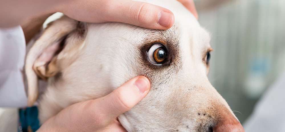
Often referred to as the eyes window, the cornea is a transparent, dome-shaped surface that serves two purposes. Along with eyelids, eyebrows, and eyelashes, the cornea aims to keep the eye free of bacteria and debris, effectively serving as a buffer between the inner eye and the environment. Due to its super sensitive nerve endings, even the gentlest touch causes an involuntary closing of the eyelid. This reflex offers protection to dogs that spend a significant amount of time outdoors or around other animals, particularly cats. The cornea also plays a key role in vision, refracting light as it enters the eye and enabling focus. Given its profound effect on vision, as well as overall quality of life, the integrity of the cornea must be preserved. If your dogs eye sustains any kind of damage, or appears to be swollen, red or irritated, immediate veterinary treatment will give your dog the best chance to retain full vision.
A dogs cornea is a three-layered powerhouse of cells that not only offers the eye a strong layer of protection, but also contributes almost two-thirds of its focusing power. Injury to the cornea must be evaluated by a veterinarian.
Vet bills can sneak up on you.
Plan ahead. Get the pawfect insurance plan for your pup.
Is There A Dog With Braces
Yes there is! Although it is not so common in Brazil, the use of canine braces is made with resin or metallic wires, correcting the position of the teeth within a period of one to four months of use, depending on the problem, says Joyce Aparecida, MD. Cobasi Corporate Education veterinarian.
The application of braces to dogs is quite uncommon, so people tend to think that this method is extremely exaggerated or unnecessary. However, the truth is quite the opposite! In the case of dogs, the braces do not have the aesthetic purpose of making their teeth prettier. In fact, it is only necessary for dogs that suffer from dental malocclusion, and it is even the best form of treatment.
Will My Dogs Eye Drops Burn When I Administer Them
Most dogs will not experience pain when you administer their eye drops.
They may squint and appear irritated in the moments after you apply them, but they should return to their normal selves within seconds of using the drops.
If you think your dog is in even more pain after applying their eye drops, we suggest reaching out to your veterinarian for guidance.
We should also note that some dogs will begin to drool after their eye drops are administered.
The tear ducts in a dogs eye will drain into the back of the nose, and in turn into their mouth in some cases.
This will cause some dogs to drool a bit due to the taste, as some of these eye drops can be extremely bitter.
If this happens, there is no need to worry.
Also Check: Ulcerative Colitis And Back Pain
Preparation Of The Platelet
PRP was prepared using a double-spin method as a protocol previously described by Kecec et al. . Briefly, blood from each animal was collected on 3.8% sodium citrate solution, soft spin at 250 à g/10 min was applied, the top and middle layers were then collected, and hard spin was performed at 2,000 à g/10 min followed by removal of platelet-poor plasma and activation of PRP by 20 mM of CaCl2 and incubation at 37°C/1 h. Centrifugation was then applied at 3,000 à g/20 min for recovering activated PRP.
Argentum Nitricum 6 30

Arg. Nit. is very useful remedy in purulent ophthalmia.
Arg. Nit. has been employed, both locally and internally, for the cure of ulceration of the cornea and fistula.
It meets old ulcerations with granular lids.
Profuse purulent discharge.
It is useful in corneal ulcer when there is great burning as if from fire.
Burning, hot discharges relieved by external warmth.
Conjunctiva looks like a piece of raw beef.
Pulsative throbbing pain in the eye.
Aggravation at night, especially after mid-night and from cold application, some amelioration from warm application.
Eyes feel very hot.
Lids are oedematously swollen and spasmodically closed.
Also Check: Ulcerative Colitis Surgery Pros And Cons
Etiology Of Corneal Ulcer
). Herpes simplex keratitis Herpes Simplex Virus Infections Herpes simplex viruses commonly cause recurrent infection affecting the skin, mouth, lips, eyes, and genitals. Common severe infections include encephalitis… read more is discussed separately.
Bacterial ulcers are most commonly due to contact lens wear and are rarely due to secondary infection from traumatic abrasion or herpes simplex keratitis. The response to the treatment depends mostly on the bacterial species, and may be particularly refractory to treatment. The time course for ulcers varies. Ulcers caused by Acanthamoeba and fungi are indolent but progressive, whereas those caused by Pseudomonas aeruginosa develop rapidly, causing deep and extensive corneal necrosis. Wearing contact lenses while sleeping or wearing inadequately disinfected contact lenses can cause corneal ulcers .
Culture And Molecular Biology
Tears samples and conjunctival swabs of all cases were streaked on mannitol salt agar and Pseudomonas agar base with CN supplement media.
Conjunctival swabs were attained by rotating a sterile cotton swab over the ventral conjunctiva and were deposited in a 2-ml tube containing sterile 0.9% NaCl solution. PCR was conducted to detect Chlamydophila felis and FHV-1 according to methods described before primers and expected amplicons are tabulated in Table 1.
Table 1. Primer sequences for Chlamydophila felis and FHV-1.
Recommended Reading: Foam Dressing For Pressure Ulcer
Treatment #: Natural Treatments
Apart from conventional and topical treatment for corneal ulcer, there are also some natural treatments that you can try for corneal ulcer treatment at home.
#1: Eye Care
When it comes to treating corneal ulcer, several treatments options are available but one of the best natural treatments you can try is to wear sunglass during the healing process. You should also avoid any kind of unnecessary eye strain. If you want to provide more comfort to your eyes then you can make use of moist warm compress on the eyes. For this, you can mix 3 cups of warm water with only 10 drops of oregano oil. After this, you need to soak a clean washcloth and then ring well and directly place over your eyes for at least 20 minutes.
#2: Try Vitamin D
When you fight against any type of eye infection, it is quite important to increase the vitamin D level in the body. However, if you experience any injury or a corneal ulcer then deficiency of vitamin D may make you more prone to develop eye infections.
When it comes to taking a high-quality vitamin D3 supplement, you should get a minimum of 10 to 15 minutes each day of direct sunlight without applying sunscreen. But, dont forget to wear sunglasses to protect your eyes. Vitamin D is proven to improve the immune system and concentration response.
#3: Dietary Changes
#4: Colloidal Silver
#5: Echinacea
My Dog Began To Drool Excessively And Paw At Its Mouth After I Administered The Eye Medications Is That A Side Effect
Not necessarily. The tear ducts drain tears away from the eyes into the back of the nose, where they then drain into the throat. Eye medications may drain through the tear ducts and eventually get to the throat, where they are tasted.
“Atropine and many other ophthalmic medications have a very bitter taste, which may cause drooling…”
Atropine and many other ophthalmic medications have a very bitter taste, which may cause drooling and pawing at the mouth. You are seeing your dog’s response to a bad taste, not a drug reaction.
Recommended Reading: Medication For Ulcerative Colitis Flare Up
Are There Any Side Effects From The Eye Medications
Occasionally a dog will be sensitive to an ophthalmic antibiotic. If your dog seems to be in more pain after the medication is used, discontinue it and contact your veterinarian immediately.
A corneal ulcer is extremely painful so the eye is usually kept tightly shut. Atropine relieves the pain but also dilates the pupil widely. Therefore, the eye is very sensitive to light and many dogs will squint or close the eye when exposed to bright light. The dilation of the eyes may last for several days after the drug is discontinued.
Identification Of Ulcer Types Treatment Plans And Complications
For superficial ulcers, a loss of part of the epithelium was the base of categorization. Deep ulcers that spread into/through the stroma and might cause severe scarring fluorescein stain was taken up by exposed corneal stroma and with green appearance. Fluorescein stain defined the corneal ulcer margin and revealed further details of the surrounding epithelium. Fluorescein dye test was applied in all the cases and used to identify the different sites of the corneal ulcer and their size.
After the ulcer type was identified, two treatment options were done: subconjunctival injection of PRP and, in case of entropion, surgical correction of entropion in affected cases followed by subconjunctival injection of PRP. Surgical correction was done under general anesthesia, as follows: atropine sulfate and xylazine were used as pre-medication, followed by ketamine at 10â20 mg/kg b.wt. for induction and maintenance .
Subconjunctival injection of autologous PRP was used in each case, and bilaterally if the bilateral ulcer was identified at the time of admission.
Subconjunctival injection of autologous PRP was scheduled and timed for each case the injection was applied under complete aseptic conditions and repeated till a complete curative clinical response with 1-week interval . Clinical evaluation was conducted via digital photographs of the ulcer at the baseline and at each time of subconjunctival injection .
Table 2. Patient signalments and description.
Read Also: Ulcerative Colitis Is It Deadly
Crystalline Deposits Causing Corneal Ulceration
There are numerous causes for the development of crystalline deposits in the canine cornea. Many do not cause ocular morbidity in terms of either pain or vision loss. Two exceptions to this general rule are corneal dystrophy in Shetland Sheepdogs, and calcareous corneal degeneration in geriatric dogs. Both of these conditions have the potential for areas of the crystalline deposits to slough and thereby cause ulcerations of the cornea. Dogs that you suspect of having either condition should be watched closely for signs of pain. I have seen several cases of calcareous degeneration in older dogs lead to stromal ulcers and perforations, so early recognition is crucial. There is no universally recognized treatment that is effective, but some cases will respond to treatment with 1% EDTA ointment in artificial tears to chelate the mineral component, or tacrolimus preparations to improve tear film quality.
What Causes A Corneal Ulcer

You May Like: What To Eat When You Have Ulcers And Acid Reflux
Corneal Abrasions Vs Corneal Ulcers
When discussing corneal injuries in dogs, you may hear the terms abrasion and ulcer used to describe the severity of the injury.
Though some may use these terms interchangeably, they are vastly different when discussing the severity of the wound.
A corneal abrasion is not as severe as a corneal ulcer, and typically involves a superficial scrape to the outermost layer of the cornea.
An abrasion has not yet eroded into the stroma, which in turn sets it apart from an actual ulcer.
Though an abrasion may not be as severe, they should still be taken seriously and treated with immediate care.
Abrasions can easily worsen without appropriate treatment, leading to the development of a corneal ulcer in the end.
Diagnosing Corneal Ulcers In Dogs
The best way to diagnose a corneal ulcer in dogs is by having your veterinarian perform a fluorescein stain of the eye.
This involves administering a bright green stain to the eye, which in turn causes the stain to pool in an ulcer if one is present.
When the veterinarian turns off the light and shines a UV light on the eye, the ulcer should become visible.
Your veterinarian should also perform an in depth eye exam to help them rule out the presence of other eye conditions.
Your vet may also use a tonometer to test the pressure in your dogs eye, as well as performing a Schirmer tear test if they think dry eye is the cause of their ulcer.
Every case will vary, so we suggest following your vets guidance when determining the best diagnostic plan.
Also Check: Nursing Care Plan For Pressure Ulcer Prevention
Corneal Ulcer Risk Factors
People who wear contact lenses are more likely to get corneal ulcers. This risk is 10 times higher if you use extended-wear soft contacts.
Bacteria on the lens or in your cleaning solution could get trapped under the lens. Wearing lenses for long periods can also block oxygen to your cornea, raising the chances of infection.
Scratches on the edge of your contact might scrape your cornea and leave it more open to bacterial infections. Tiny particles of dirt trapped under the contact could also scratch your cornea.
Other things that may make you more likely to have a corneal ulcer include:
- Steroid eye drops
- Eyelashes that grow inward
- Eyelids that turn inward
- Conditions that affect your eyelid and keep it from closing all the way, like Bellâs palsy
- Chemical burns or other cornea injuries
What Is A Corneal Ulcer
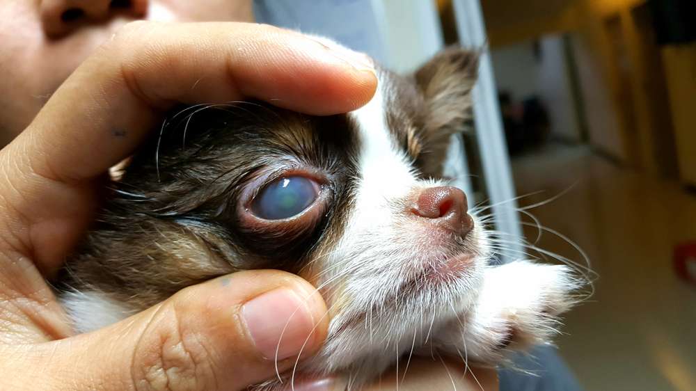
Erosion of a few layers of the epithelium is called a corneal erosion or corneal abrasion. A corneal ulcer is deeper erosion through the entire epithelium and into the stroma. With a corneal ulcer, fluid accumulates in the stroma, giving a cloudy appearance to the eye. If the erosion goes through the epithelium and stroma to the deepest level of Descemet’s membrane, a descemetocele is formed. A descemetocele is a very serious condition. If Descemet’s membrane ruptures, the liquid inside the eyeball leaks out, the eye collapses and irreparable damage occurs.
Recommended Reading: Can Stress Cause Flare Up Ulcerative Colitis
Corneal Ulcers In Dogs
Eye issues are taken very seriously in veterinary medicine. When we hear that a pets eye is squinting, red, swollen, irritated, appears glossy, or is producing discharge, we encourage pet owners to go directly to a veterinary hospital to have it assessed. Sometimes, it is just a simple eye infection which can be treated with antibiotics.
But sometimes, it can be a more serious underlying disease or injury such as glaucoma, cataracts, or corneal ulcers. These more advanced eye issues progress quickly and if gone untreated, they can cause serious damage or blindness. Corneal ulcers are particularly common in small animal medicine and depending on how deep it is, can be very dangerous for your pet.
The Cornea: The Window of the Eye
The cornea is the transparent window that makes up the front surface of the eye. It is composed of three main layers:
Corneal Ulcers: Sometimes Serious, Sometimes Not
A corneal ulcer is a wound or abrasion on the corneal surface. A superficial corneal ulcer involves only the surface epithelium. This is a much less serious injury, but still requires veterinary care.
Causes: Most Commonly Trauma
- A cat scratch or a sharp object
How To Diagnose Corneal Ulcer
Diagnosis of a corneal ulcer is very important in the early stage so that one can easily treat corneal ulcer with ease. During the diagnosis, your doctor will ask some questions so that he/she can determine the cause of the ulcers. There will be a great need to examine the eye under a bio-microscope, known as a slit lamp. This exam will help the doctor to see the damage to the cornea and then determine if you have a corneal ulcer. For this, fluorescein, a special dye will be placed into the eye to light up the area and helps in the diagnosis process.
However, if the exact cause of the corneal ulcers is still unknown then your doctor will take a small sample of the ulcer to know how it can be treated properly. After your eyes are numbed with the eye drops, the cells may be scraped gently from the corneal surface so that the ulcer can be easily tested.
Read Also: How To Cure Ulcerative Proctitis
What Are Risk Factors For A Corneal Ulcer
The following are risk factors for corneal ulcers.
- The number one risk factor for corneal ulcer in the U.S. is contact lens wear.
- Trauma to the eyes
- Feeling that there is something in your eye
- Obvious discharge draining from your eye
- If you have a recent history of scratches to the eye or exposure to chemicals or flying particles
Alternative Medicine For Corneal Ulcer Treatment

Use of contact lenses increases your risk of a corneal ulcer, as would a chronic dry eye condition, so taking a break from contact lenses and finding a way to improve eye moisture levels is a great start. Some people have found castor oil to be beneficial to soothe and moisturize dry eyes. Supplemental Vitamin C, Vitamin A, and protein may help repair damage from a corneal ulcer and prevent a corneal ulcer scar.
Also Check: Pepto Bismol And Ulcerative Colitis
Surgical Treatment Of Canine And Feline Descemetoceles Deep And Perforated Corneal Ulcers With Autologous Buccal Mucous Membrane Grafts
Oculistica Veterinaria Genova, Genova, Italy
Department of Veterinary Science, University of Pisa, Pisa, Italy
Department of Veterinary Science, University of Pisa, Pisa, Italy
Correspondence
Samanta Nardi, Department of Veterinary Science University of Pisa, via Livornese lato monte, 56122, San Piero a Grado, Pisa, Italy.